Ingela Nyström's Early Career Research
Collaboration partners:
-
Professor Gunilla Borgefors
Centre for Image Analysis
-
Dr. Gabriella Sanniti di Baja
Istituto di Cibernetica,
National Research Council of Italy (CNR), Pozzuoli (Napoli), Italy
Dr. Sanniti di Baja promoted as honorary doctor at Uppsala University, 2002
-
Professor Örjan Smedby
Dept. of Radiology, University Hospital, Linköping
-
Dr. Stina Svensson
Centre for Image Analysis
-
Professor Jayaram K. Udupa
Medical Image Processing Group, Dept. of Radiology, University of Pennsylvania, Philadelphia, USA
Projects:
-
Area of and Volume Enclosed by Digital and Triangulated Surfaces
-
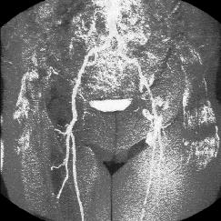
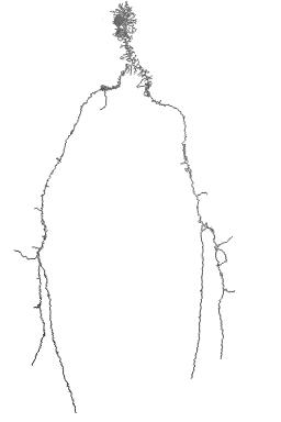
Skeletonization applied to magnetic resonance angiography images.
Left: Maximum intensity projection of an MRA image of the arteries of the pelvis.
Right: The corresponding (pruned) curve skeleton of the segmented blood vessels.
-
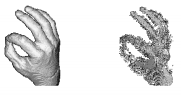
Reducing objects to "skeletons" in the process of quantifying its shape.
Toriwaki/Katada hand.

-
Filling concavities for quantifying the shape of an object.

-
Different applications for the distance transform.
Above the 3-4-5 sphere.