Follow this link for online registration.
Registration closes Friday June, 9th. 1-day passes are available.
Travel Grants
Travel grants are available for PhD students, Post Docs and research engineers through SCANDEM. Apply through the SCANDEM homepage
(Grants – SCANDEM) with deadline on the 30th of April.
Payment instructions
Swedish participants
Swedish participants can choose to pay either by credit card or by invoice. Invoices to Swedish organizations will be sent as an electronic invoice (e-faktura) if possible.
Participants from outside Sweden
All payments must be made in SEK (Swedish krona). Payment should be made by credit card or by bank transfer (invoice).
Invoice
When choosing ‘International bank transfer’ in the registration form, you will receive an invoice including bank transfer information (bank account number, IBAN, etc). The invoice is created manually, and will be sent as a pdf attachment by e-mail, usually a few days after registration. The participant/payee is responsible for any bank charges/fees associated with the payment.
Information about payment with credit card
We accept the following credit cards: VISA and Mastercard.
For your safety, our payment system is connected to the 3D Secure system for ensuring the security of payments made through the internet. Your card needs to be connected to Verified by Visa or Mastercard SecureCode. Your bank can inform you if your card is connected to one of these advanced security payment programs, and if not already, how it can be connected
VAT
According to Swedish and EU legislation, 25% Swedish VAT will be added to all registration fees.
This according to EU legislation and regulation for conferences and events (C-647/17, EU:C:2019:195, of 13 March 2019 and Article 53 of Council Directive 2006/112/EC of 28 November 2016) VAT is paid in the country where services are rendered, e.g. the country in which the conference takes place.
Exception: Swedish participants from governmental public sector organizations (e.g., universities, governmental agencies): No VAT will be added. In this case, you must choose Invoice (Swedish billing address only) as the payment mode and provide the VAT number of your employer.
Cancellation of Registration
Notification of cancellation must be made in writing and sent to the conference secretariat.
Cancellation of registration will be accepted until 14 May 2023 up to which date the total amount will be refunded except for a cancellation fee of 750 SEK. We regret that no refunds or reductions of fees will be made for notification of cancellations received after 14 May 2023, nor for no-shows for any reason.
Change of Name
Should you be unable to attend, a colleague can take your place. Please contact the conference secretariat as soon as possible.
Conference secretariat e-mail:scandem2023@akademikonferens.se
GDPR
Academic Conferences, in-house Professional Congress Organizer of Karolinska Institutet, SLU, Stockholm University and Uppsala University, is the administration office of SCANDEM 2023.
We administer conferences and other events. In order to do this, we must collect and process personal data. The purpose of our privacy policy is to explain the data we collect and how we use it, as well as your rights as a data subject.
SCANDEM2023 welcomes submissions of abstracts presenting original research on microscopy instrumentation, techniques and applications within material and life science. Submission closes March 31 April 16 and notice of acceptance is April 30. Late abstracts for poster presentation still accepted! Prepare your abstract according to this word template or pdf instruction/template
Submit your abstract via our on-line system using this link.
Abstract submission is now closed.
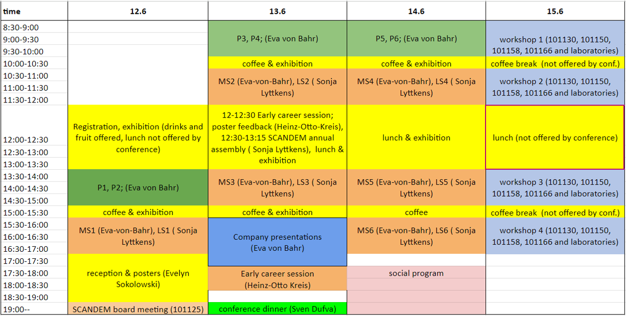
An overview of the program is shown below. More detailed information about the program and events can be found in the
program booklet and all abstracts are found in the
abstract book . The plenary sessions each contain two plenary talks of length 45 min. Short bios of the plenary speakers as well as titles and abstracts for their talks are presented below. The parallel sessions will have tracks with material and life science focus, respectively. The invited talk in each session is 30 min including questions. Short bios and abstracts of the invited session talks are presented below. The contributed (abstract submitted for oral presentations) material science talks are 15min including questions and the contributed life science talks are 20 min including questions.
Program overview
Session topics:
Material science
- MS1. Refinement and development of microscopy techniques
- MS2. Studies for structure and strain in materials
- MS3. Electron microscopy in-situ and in-operando techniques
- MS4. Functional materials
- MS5. Microscopy of soft and beam-sensitive materials
- MS6. Use of spectroscopic techniques in material science and geoscience
Life science
- LS1. Image analysis and data visualization
- LS2. Beyond the resolution revolution - new challenges in cryo-EM/ET
- LS3. Correlative light and electron microscopy
- LS4. Ultrastrucutral pathology and disease understanding
- LS5. Automated and AI based microscopy imaging and analysis
- LS6. In situ and live cell microscopy
Poster session
The boards for posters are of size: width 1m, height 1.3m. Posters can be mounted from Monday 11:00 and should be unmounted latest
on Tuesday 17:30.
Early career session
This session of the conference is dedicated to developing scientific visualization skills and aimed at early career researchers, but, of course, open for any participant to join free of charge. To realize this goal, we have engaged the services of Andreas Dahlin and his company "Visualize your Science" to realize two parts of the program:
- Tuesday lunch break: poster feedback in small groups under professional guidance
- Tuesday 17:30-18:30: how-to presentation on poster design/scientific visualization
About Andreas Dahlin and "Visualize your Science":
Andreas has experience from the industry and academia, having worked in pharma for three years and becoming Assoc. Prof. at Uppsala University in 2017. During his time at Uppsala, Andreas got known as the go-to guy for illustrations and started the Visualize Your Science course in 2011 due to popular demand. As the interest in the course grew, he saw an opportunity to teach full-time and left academia in 2016 to start to Visualize your Science AB. Today there are five visual scientists in the company, giving courses to around 600 PhD students yearly in visual communication skills for scientists.
Social program
Information about the social program can be found in the
program booklet.
Company presentation session
Information about which companies that will introduce themselves can be found in the
program booklet.
Plenary speakers
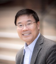
- P1. Xiaoqing Pan
-
University of California, Irvine, USA
-
Emergent Phonon Phenomena at Interfaces Probed by Vibrational Electron Microscopy
-
Abstract: Crystal defects and interfaces affect the thermal and heat-transport properties of materials by scattering phonons and modifying phonon spectra. Spatially resolved vibrational mapping of nanostructures and defects is indispensable to the development and understanding of thermal nanodevices, modulation of thermal transport and novel nanostructured thermoelectric materials. Through the engineering of complex structures, such as alloys, nanostructures and superlattice interfaces, one can significantly alter the propagation of phonons and suppress material thermal conductivity while maintaining electrical conductivity. There have been no correlative experiments that spatially track the modulation of phonon properties in and around individual defects and nanostructures due to spatial resolution limitations of conventional optical phonon detection techniques. In this talk, we demonstrate that space- and angle-resolved vibrational spectroscopy in a transmission electron microscope makes it possible to map the vibrational spectra of a single interface and a quantum dot. We detect a red shift of several meV in the energy of acoustic vibration modes near a single stacking fault in cubic silicon carbide, together with substantial changes in their intensity, and find that these changes are confined to within a few nanometers of the stacking fault. In a two-dimensional lateral heterostructure, new phonon modes at 27.9 and 41.1 meV are observed at the interface between monolayer thick MoS2 and WSe2. At a Si-Ge interface, localized interfacial phonon modes at ~48 meV. Simulations show that these interfacial phonon modes contribute to the total thermal interface conductance. By tracking the variation of the Si optical mode in a phonon map from a single SiGe quantum dot, the nanoscale modification of the composition-induced red shift is observed. In this work we have also developed a novel technique to differentially map phonon momenta, providing direct evidence that the interplay between diffuse and specular reflection largely depends on the detailed atomistic structure. Our work unveils the non-equilibrium phonon dynamics at nanoscale interfaces and can be used to study actual nanodevices and aid in the understanding of heat dissipation near nanoscale hotspots, which is crucial for future high-performance nanoelectronics.
-
Short bio: Xiaoqing Pan is an internationally renowned materials scientist and leading expert in electron microscopy with his pioneering development and applications of novel transmission electron microscopy (TEM) methods for understanding the atomic scale structure, properties and dynamic behaviors of functional materials. His research has led to the development of new materials with novel functionalities and discoveries of new phenomena materials. He has published over 400 peer-reviewed scientific papers in high impact factor journals such as Nature, Science, and Nature Materials. His publications have been highly cited, and he is recognized as one of the Clarivate’s highly cited researchers in 2020. He has given over 230 invited talks (including keynote and plenary presentations) at national and international scientific conferences and at more than 200 invited seminars at other institutions. He received the National Science Foundation’s CAREER Award and the China Natural Science Foundation’s Outstanding Young Investigator Award. He is a Fellow of the American Ceramic Society, American Physical Society, Microscopy Society of America, and the Materials Research Society. Pan is currently the Henry Samueli Endowed Chair in Engineering, Professor of Materials Science and Engineering, and Professor of Physics & Astronomy at UCI. In addition, he is the founding Director of the Irvine Materials Research Institute (IMRI), and the founding Director of the Center for Complex Active Materials (CCAM – an NSF MRSEC).
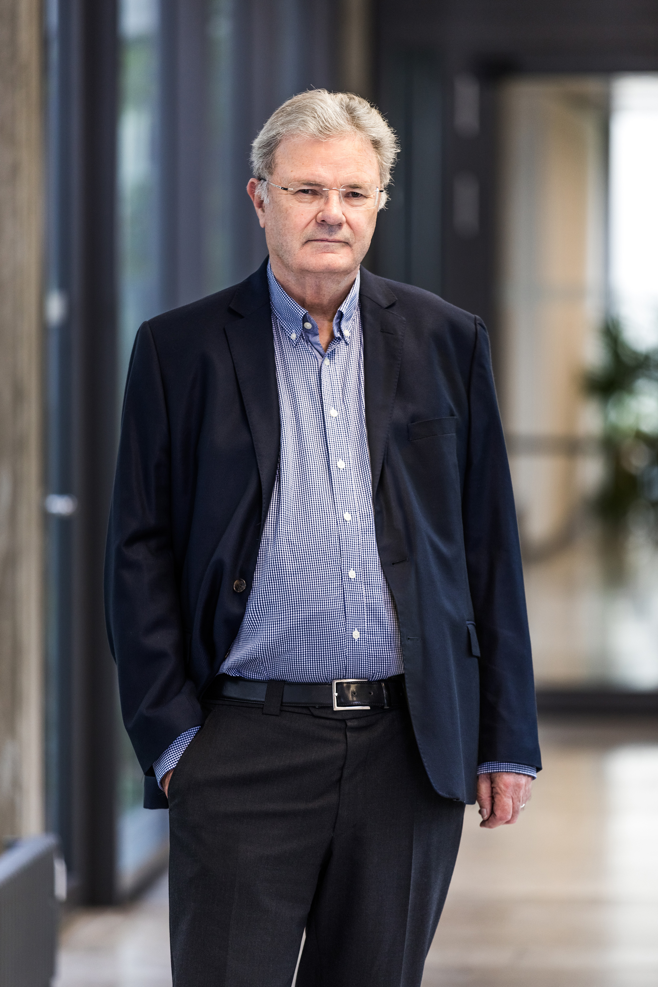
- P2. Wolfgang Baumeister
-
Max Planck Institute of Biochemistry, Am Klopferspitz 18, 82152 Martinsried, Germany
-
Cryo-Electron Tomography or the Power of Seeing the Whole Picture
-
Abstract:Traditionally, structural biologists have approached cellular complexity in a reductionist manner by studying isolated and purified molecular components. This 'divide and conquer' approach has been highly successful, as evidenced by the impressive number of entries in the PDB.
However, awareness has grown in recent years that only rarely can biological functions be attributed to individual macromolecules. Most cellular functions arise from their acting in concert. Hence there is a need for methods developments enabling studies performed in situ, i.e. in unperturbed cellular environments. Sensu stricto the term 'structural biology in situ' should apply only to a scenario in which the cellular environment is preserved in its entirety.
Cryo electron tomography has unique potential to study the supramolecular architecture or 'molecular sociology' of cells. It combines the power of three-dimensional imaging with the best structural preservation that is physically possible to achieve. We have used this method to study the 26S proteasome in a number of cellular settings revealing their precise location, assembly and activity status as well as their interactions with other molecular players of the cellular protein quality control machinery.
-
Short bio: Wolfgang Baumeister obtained his PhD from the University of Düsseldorf in 1973. 1981/82 he spent time at the Cavendish Laboratory in Cambridge, England as a Heisenberg Fellow. In 1982 he joined the Max-Planck-Institute of Biochemistry in Martinsried as a group leader. Since 1988 he is a Scientific Member of the MPG and Director of the Department of Structural Biology. He is a Honorary Professor at the Technical University of Munich in the departments of Physics and Chemistry. Since 2018 he is also a Distinguished Professor at ShanghaiTech University.
Professor Baumeister’s main interest is the development of new tools and methods for the structural characterization of molecules and cells. He is particularly interested in the development of cryo-electron tomography for structural studies of molecular and supramolecular structures in situ, i. e. in their native cellular habitats. A major focus of his work is the molecular machinery of protein degradation, in particular via the ubiquitin-proteasome pathway, and autophagy. He is a member of the German Academy of Sciences, Leopoldina, the US National Academy of Sciences and the American Academy of Arts and Sciences.
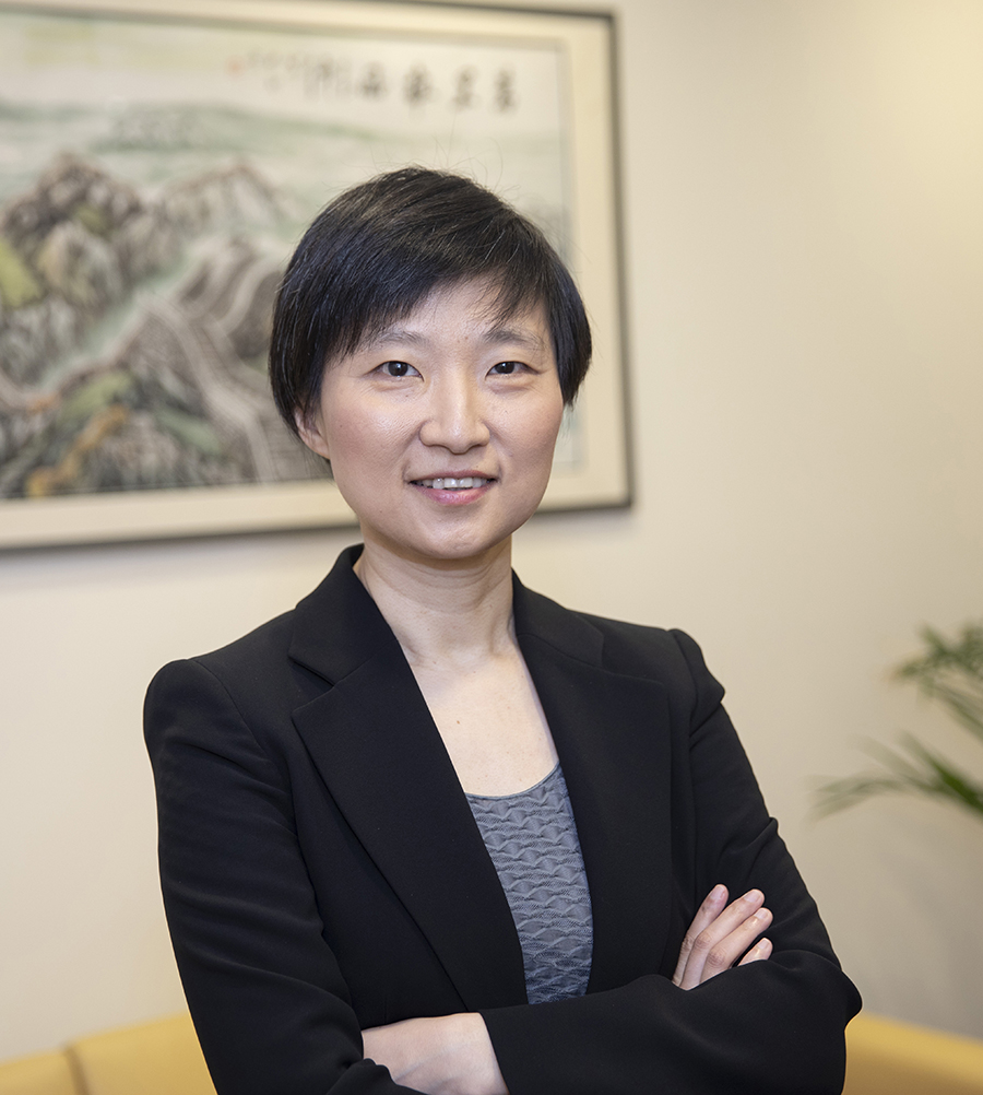
- P3. Xiaowei Zhuang
-
Harvard University, Howard Hughes Medical Institute, USA
-
Spatially resolved single-cell genomics & cell atlas of the brain
-
Abstract: Inside living organisms, thousands of different genes function collectively to give rise to cellular behavior and tissue function. Understanding the behaviors and functions of cells and tissues thus require imaging at the genome scale, which will advance our understanding in many areas of biology, ranging from the regulation of gene expression in cells to the development of cell fate and the organization of cell types in complex tissues. We developed a single-cell transcriptome and genome imaging method, multiplexed error-robust fluorescence in situ hybridization (MERFISH), which allows RNA, DNA, and epigenetic marks to be imaged at the genome scale. This approach enabled spatially resolved transcriptomic profiling, epigenomic profiling, and 3D-genome organization mapping in single cells. The ability to perform single-cell gene expression profiling in intact tissues further enabled the identification, spatial mapping, and functional annotation of distinct cell types in intact tissues. In this talk, I will describe the MERFISH technology and its applications, with a focus on mapping the molecular, spatial, and functional organizations of cell types in the mouse and human brain.
-
Short bio: Xiaowei Zhuang is an investigator of Howard Hughes Medical Institute and the David B. Arnold Professor of Science at Harvard University. She pioneered the development of super-resolution imaging and genome-scale imaging methods. She invented STORM, a super-resolution imaging method, and discovered novel molecular structures in cells using STORM. She invented a single-cell transcriptome and genome imaging method, MERFISH, and made discoveries in the areas ranging from the 3D genome organization and gene regulation in cells to the cellular organization and functions in the brain using MERFISH.
Zhuang received her B.Sc. degree in physics from the University of Science and Technology of China, her Ph.D. in physics under the supervision of Prof. Y. R. Shen from University of California at Berkeley, and her postdoctoral training in biophysics in the lab of Prof. Steven Chu at Stanford University. She joined the faculty of Harvard University in 2001 and became a Howard Hughes Medical Institute investigator in 2005.
She is a member of the National Academy of Sciences, National Academy of Medicine, and the American Academy of Arts and Sciences, a fellow of the National Academy of Inventors, a member of the American Philosophical Society, and a foreign associate of the Chinese Academy of Sciences and the European Molecular Biology Organization. She received honorary doctorate degrees from the Stockholm University, the Delft University of Technology, and the Icahn School of Medicine at Mount Sinai. She has received many awards, including the Heinrich Wieland Prize, J. Allyn Taylor International Prize in Medicine, the FNIH Lurie Prize in Biomedical Sciences, the Vilcek Prize in Biomedical Science, the Breakthrough Prize in Life Sciences, the Pearl Meister Greengard Prize, the National Academy of Sciences Award for Scientific Discovery, the Heineken Prize for Biochemistry and Biophysics, the National Academy of Sciences Award in Molecular Biology, the Raymond and Beverly Sackler International Prize in Biophysics, the Max Delbruck Prize in Biological Physics, the MacArthur Fellowship, etc.
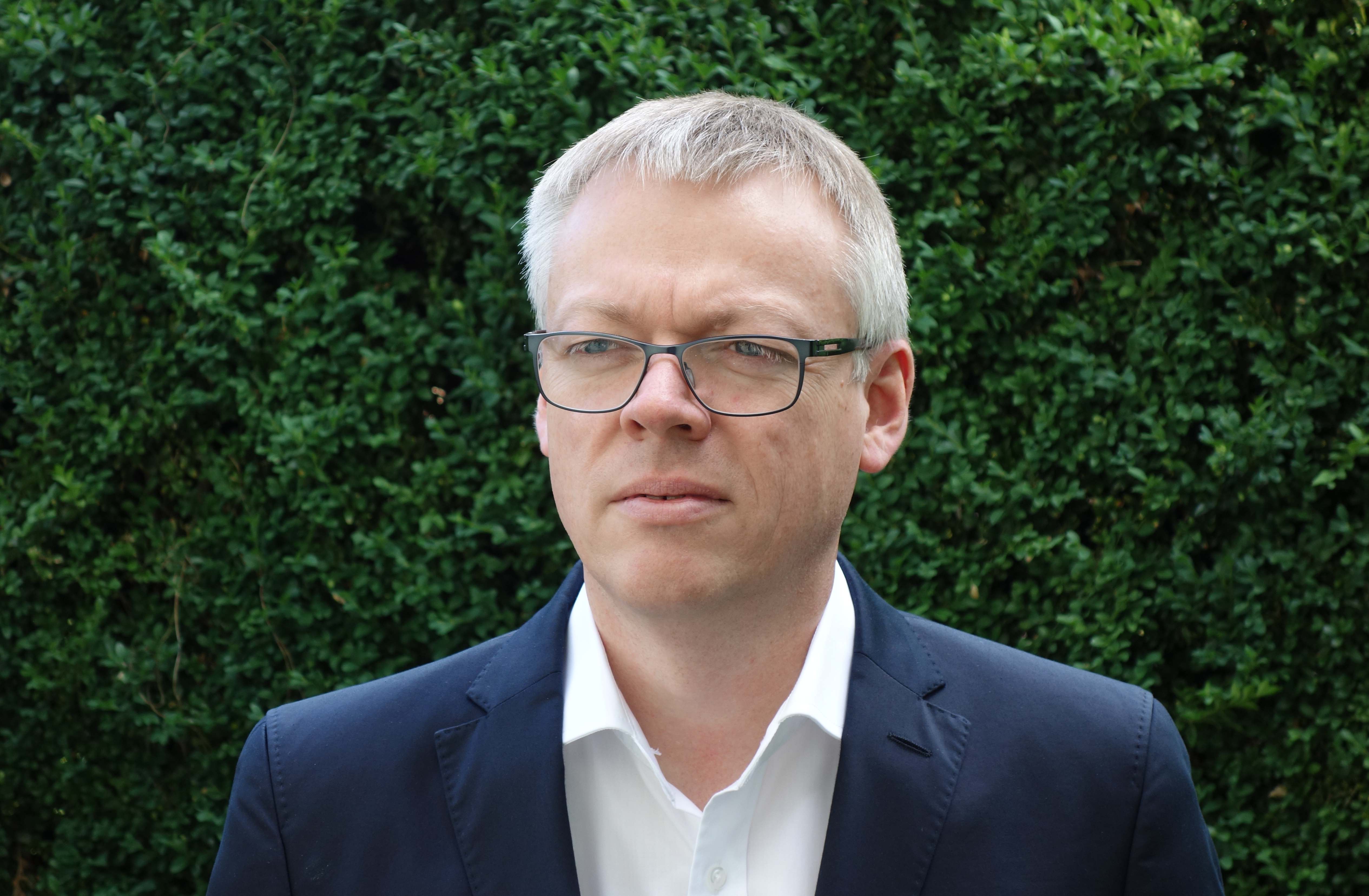
- P4. Michael Laue
-
Robert Koch Institute, Berlin, Germany
-
Electron microscopy in infectious diseases – a medical perspective
-
Abstract: Exciting developments in light and electron microscopy over the past decades have provided better insights and literally pictures of how pathogens interact with cells at the cellular, subcellular and molecular level. However, the reaction which a pathogen causes in the human body is complex and goes beyond the interaction of the pathogen and the individual cell. Usually, it is a systemic reaction which involves a complex interplay between several systems of the human body. The presentation will highlight this aspect by showing examples from studying COVID-19 disease and the recent outbreak of the mpox. A side view will discuss the strengths and weaknesses of infectious disease models. Finally, an outlook tries to define which developments are necessary in (electron) microscopy for improving the research of infectious diseases in future.
-
Short bio: Michael Laue studied biology and mainly used electron microscopy for investigating research questions in various fields of biology and medicine. After working for the Max-Planck Society and several medical faculties in Germany, he became head of the microscopy division at the Robert Koch Institute, which is the main public health institute in Germany. Besides studying how microorganisms live and perform, the microscopy division supports the public health sector with diagnostic electron microscopy in infectious diseases.
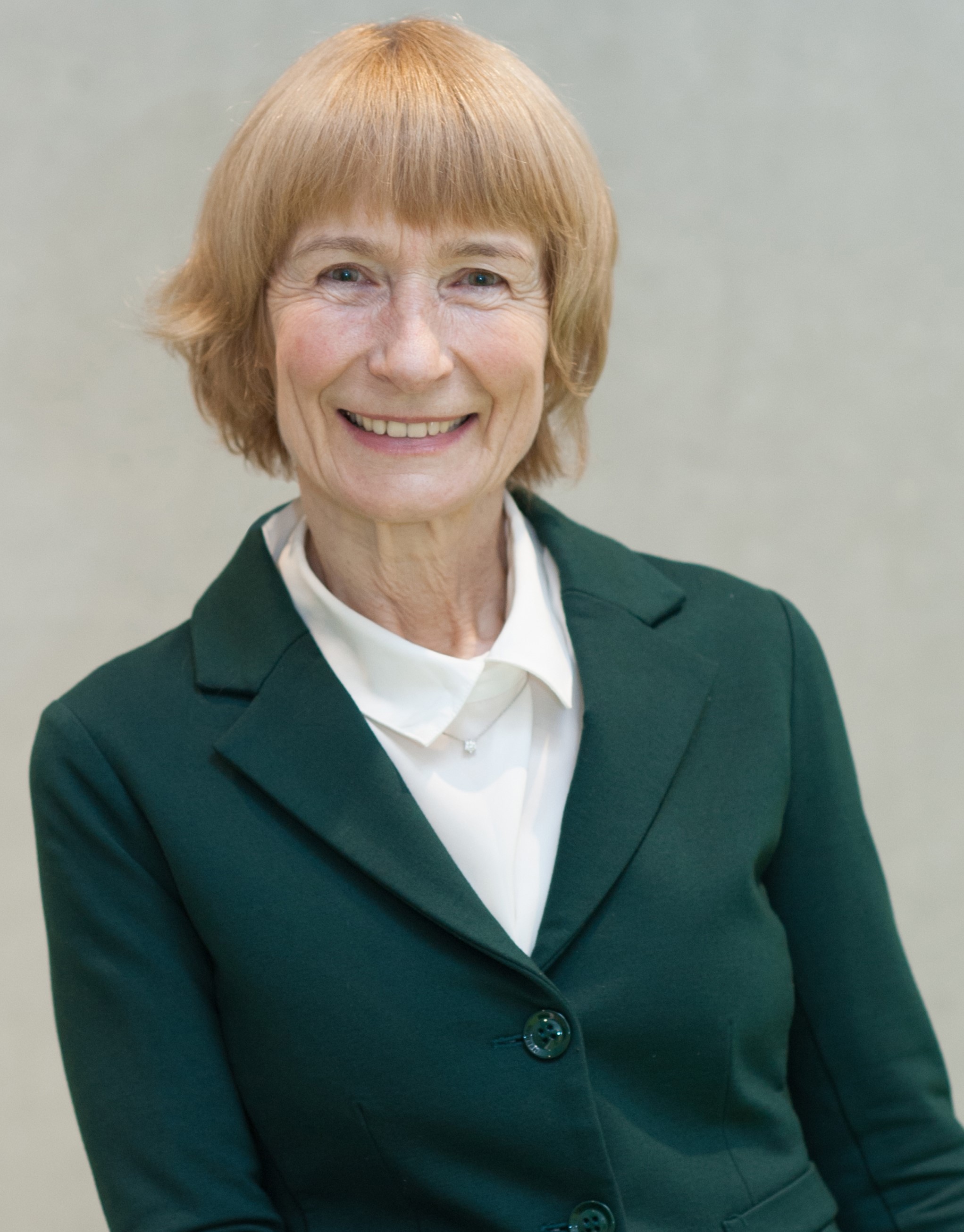
- P5. Ute Kaiser
-
Ulm University, Germany
-
Findings from the happy marriage between low-dimensional materials and low-voltage atomically-resolved TEM
-
Abstract: We have known for about 2500 years that the world is made of atoms. But only recently have we learned that the well-defined arrangement of atoms in two dimensions leads to materials with extraordinary solid-state and quantum properties. And it is precisely in these well-defined thin materials that we are beginning to use the developments in aberration-corrected low-voltage electron microscopy that have taken place in parallel to image individual defects including their movement in these thinnest materials and to modify the material in a targeted manner. In the lecture we show selected examples.
-
Short bio: Ute Kaiser received her diploma, her PhD in crystallography from the Humboldt University Berlin, and her habilitation in experimental physics from the Friedrich-Schiller University, Jena, Germany, in 2002. Since 2004 she is full professor at Ulm University in the Physics Department and Head of Ulm’s Materials Science Electron Microscopy Centre. Her research interests comprise the areas of TEM methods and instrumentation development, and applications in the fields of battery, semiconductor, and catalysts materials. From 2009 till 2018 she was the scientific director of the SALVE (Sub Angstroem Low-Voltage Electron Microscopy) project to develop low-voltage transmission electron microscopy, including the development of the chromatic and spherical aberration-corrected instrument, sample preparation methods and contrast calculation at lower accelerating voltage. By means of this unique TEM instrument, she dedicates her work to unravel the crystallographic and electronic properties of low-dimensional materials. Ute Kaiser has more than 400 peer-reviewed articles, and holds several honorary adjunct positions. She is currently the Physical-Sciences Editor for the Journal Micron.
- P6. Frances M. Ross
-
Massachusetts Institute of Technology in Cambridge, MA, USA
-
In situ TEM techniques for controlling structure and interfaces in 2D material
-
Abstract: The properties of two dimensional (2D) van der Waals materials are remarkably varied, but can be expanded even further if we pattern the layers in-plane or create well-defined interfaces between the 2D layer and other types of materials. In situ electron microscopy helps develop and refine these strategies by providing spatially and temporally resolved information while we perform these modifications. I will first discuss interfacial control, showing how in situ deposition and annealing experiments in an ultra-high vacuum TEM enable quantification of the nucleation and growth of epitaxial nanocrystals and more complex, multi-component nanostructures on 2D surfaces. I will then discuss patterning, using 2D magnets as an example. Here, electron beam-induced atomic displacements, controlled through machine learning techniques, enable the structure to be modified at individual lattice sites, potentially tailoring the magnetic texture. Given the variety of fascinating materials problems and the exciting advances in instrumentation and data analysis that are ongoing, it seems likely that in situ techniques will continue to play their unique role in the development of new structures and functionalities based on 2D materials.
-
Short bio: Frances M. Ross is at the Department of Materials Science and Engineering at the Massachusetts Institute of Technology in Cambridge, MA, USA. She received her B.A. in Physics and Ph.D. in Materials Science from Cambridge University, UK, where she became captivated by electron microscopy. She continued this interest during her postdoc at A.T.&T. Bell Laboratories, as a Staff Scientist at the National Center for Electron Microscopy, Lawrence Berkeley National Laboratory, and as a Research Staff Member at the IBM T. J. Watson Research Center. She joined MIT in 2018. Her research is based around the development of in situ electron microscopy techniques to help understand crystal growth, epitaxy, self-assembly and electrochemical and other liquid phase processes.
Invited session speakers
- MS1. Knut Müller-Caspar
-
Ludwig-Maximilians-Universität Münch, Munich, Germany
-
Multidimensional TEM – characterization of electric fields and structure based on full momentum resolution
-
Abstract: In recent years, the dimensionality in transmission electron microscopy (TEM) has increased rapidly by the advent of ultrafast cameras that record at frame rates of many kHz. This development has especially paved the way for a revolution as to the versatility of scanning TEM (STEM). In particular, momentum-resolved STEM enhanced traditional Z- and phase-contrast techniques such that any conventional imaging mode is present simultaneously in a 4D data set. Most importantly, the combination of real- and reciprocal space information nowadays allows to quantify charge densities with subatomic resolution, to measure polarisation-induced electric fields, and to solve the phase problem by ptychographic techniques. This presentation demonstrates the capability of 4D-STEM by means of several examples such as the mapping of atomic electric fields in 2D materials and ptychographic reconstructions using different algorithms. Furthermore, mapping of polarization-induced electric fields in semiconductors and ferroelectrics is addressed using GaN/AlN systems and PbZrTiO. Finally, factors impacting the quantitative interpretation of 4D STEM data are worked out, namely the role of probe focus and momentum transfers due to (multiple) plasmon scattering.
-
Short bio: Knut Müller-Caspary received his Ph.D from Bremen University (Germany) in 2011. Between 2011 and 2016 he worked as a postdoctoral research fellow in Bremen focussing on strain, composition and electric field mapping by momentum-resolved STEM. He established cooperations with several companies to explore new detectors as to speed, dynamic range, efficiency and in-situ capability. In particular, he contributed key developments to the mapping of atomic electric fields and charge densities at subatomic scale by exploiting the full complexity of STEM diffraction patterns. In 2016 Müller-Caspary moved to the EMAT institute at the University of Antwerp (Belgium) where he applied STEM to the electrical characterisation of 2D materials. In 2018 he established a Helmholtz Young Investigator Group for momentum-resolved STEM at Forschungszentrum Jülich (Germany) and became junior professor at RWTH Aachen University in 2019. In 2021, Knut Müller-Caspary moved to the faculty of chemistry and pharmacy at Ludwig-Maximilians-University Munich as university professor for TEM.
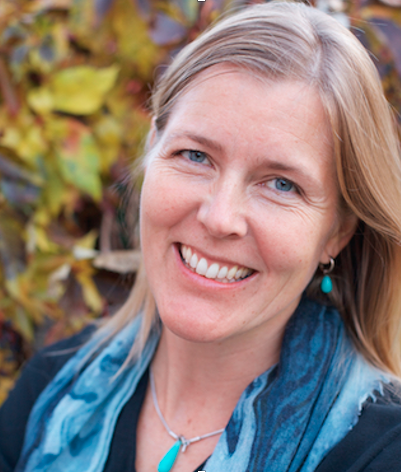
- LS1. Carolina Wählby
-
Uppsala University
-
Spatial transcriptomics
-
Abstract: Spatial transcriptomics combines microscopy with groundbreaking molecular methods enabling detection of hundreds of active genes in parallell, directly in a tissue sample at sub-cellular resolution. This opens up for new possibilities of linking gene expression to health, development and disease, and also to local tissue morphology. In my talk, I will focus on possibilities and recent results when it comes to understanding organ development and cancer, and showcase some of our latest tools for data visualization and computational analysis of this information-rich data.
-
Short bio: Carolina Wählby is professor in Quantitative Microscopy at the Dept. of Information Technology, Uppsala University, and Scientific Director of the National SciLifeLab Bioimage Informatics facility. Her research lies in the intersection between life science and computational image analysis, developing image analysis, deep learning, and visualization approaches for understanding dynamics of cancer tissue, antibiotics susceptibility and spatial transcriptomics, funded primarily by the ERC and the Swedish Foundation for Strategic research. She has a MSc in molecular biotechnology and a PhD in digital image analysis, and carried out postdoc research within genetics and pathology. She was part of the Imaging Platform of the Broad Institute 2009-2015, developing CellProfiler, and became full professor at Uppsala University in 2014. She received the SBI2 President’s innovation award in 2014, and the Thuréus prize in 2015 and is a member of the Royal Swedish Academy of Engineering Sciences. She is a member of the steering group of a 300M€ effort on Data Driven Life Science, funded by the Knut and Alice Wallenberg Foundation, with the ambitious goal of training the next generation of life scientists.
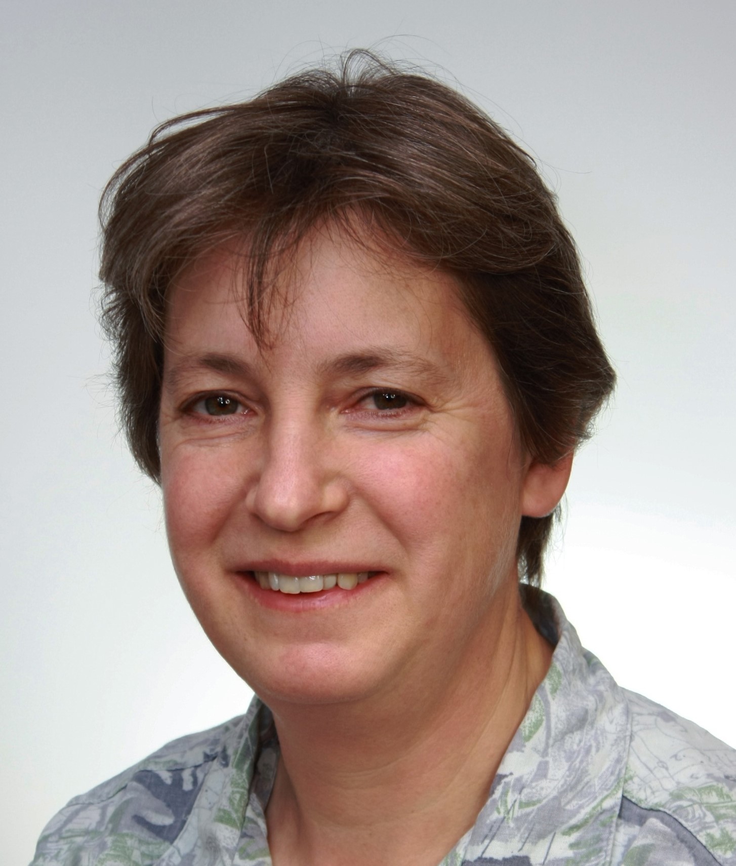
- MS2. Randi Holmestad
-
Norwegian University of Science and Technology, Trondheim
-
4DSTEM used to study Precipitates in age-hardenable Aluminium Alloys
-
Short bio: Randi Holmestad has been a professor at Department of Physics, NTNU, Trondheim, Norway since 1999. She completed her PhD in materials physics (on quantitative convergent beam electron diffraction) from the same university in 1994. Holmestad's present research interests are focused on materials physics; transmission electron diffraction and microscopy (TEM), materials microstructure and the relations to macroscopic properties. Her research group has high expertise in using advanced TEM techniques to study (aluminium) alloys, scanning TEM, and different diffraction techniques, including scanning precession electron diffraction (SPED) and convergent beam electron diffraction (CBED). Holmestad has had several sabbaticals abroad, latest at University of Illinois, Urbana-Champaign, IL, USA, in 2005 and 2012, and at Monash University, Australia for 2 months during fall 2019. She is the leader of the NORTEM infrastructure project, the TEM Gemini Centre, a program head in SFI-CASA, and SFI PhysMet and a board member in EMS. The TEM group in Trondheim is a member of ESTEEM3.
- LS2. Kay Grunewald
-
Leibniz Institute of Virology and Universität Hamburg, Germany
-
Strucutral cell biology of membrane modulations by herpesviruses
-
- MS3. Kimberly Dick Thelander
-
Lund University, Sweden
-
Real Time Investigation of Crystal Growth and Nucleation of Low-dimensional Inorganic Nanostructures
-
Abstract: A major challenge to designing new, novel nanostructures with properties tailored according to specific needs is understanding at an atomic scale the nanoscale processes and size effects that lead to structures with unique properties. To address this we use atomically-resolved in-situ TEM to follow the nanoscale crystal growth process in real time, which offers insights into the growth processes unparalleled by any other technique. Our approach uses an aberration-corrected environmental TEM connected to a chemical vapor deposition system designed for compound semiconductor growth with separate gas lines for supplying the different precursor gases. In this talk I will discuss recent investigations into the crystal growth of semiconductor nanostructures, including nanowires and nanoparticles.
-
Short bio: Kimberly Dick Thelander is a Professor of Materials Science at the Centre for Analysis and Synthesis, Department of Chemistry, Lund University, Sweden, and a member of the Royal Swedish Academy of Engineering Sciences. She received her PhD degree in Physics in 2007 from Lund University, following a Bachelor of Science in Chemical Physics in 2003 from the University of Waterloo in Canada. Her research involves the crystal growth of novel semiconductor nanomaterials, with particular focus on understanding the processes and conditions that lead to the formation of metastable crystal phases and alloys.
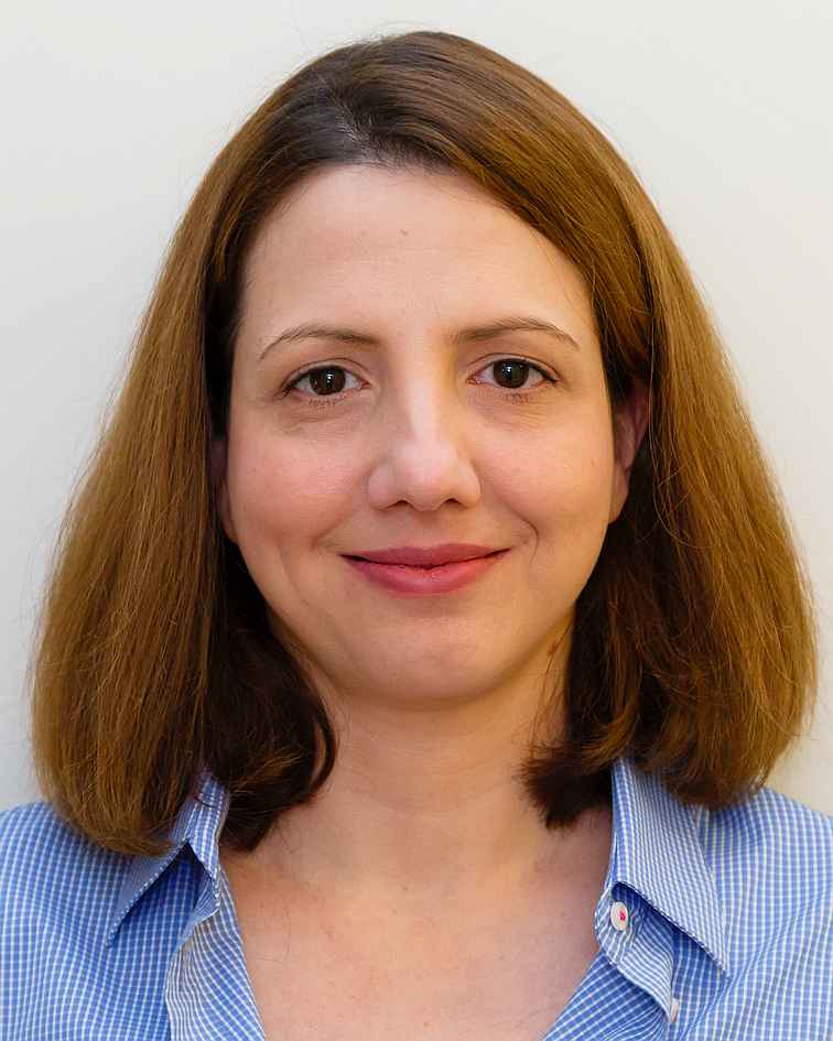
- LS3. Marie-Charlotte Domart
-
Crick Institute, UK
-
Correlative light, electron and X-ray microscopy: finding the needle in the haystack -
Abstract: Fluorescence microscopy is a powerful tool for localising proteins within biological samples. However, information is limited to the distribution of the tagged protein, telling us little about the ultrastructure of the surrounding cells and tissues, which may be intimately involved in the biological process under study. Electron microscopy overcomes the resolution limitation inherent in light microscopy and can reveal the ultrastructure of cells and tissues. However, protein localisation tends to be complex and is often dependent on the availability of ‘EM-friendly’ antibodies. Correlative light and electron microscopy (CLEM) combines the benefits of fluorescence and electron imaging, revealing protein localisation against the backdrop of cellular architecture. In this talk, I will introduce several ways in which we are developing 3D CLEM for application to biological samples.
-
Short bio: Marie-Charlotte Domart is a Principal Laboratory Research Scientist in the Electron Microscopy Science Technology Platform at the Francis Crick Institute in London, UK. Biologist by training, she has a MSc in Oncology and a PhD in Biochemistry and Molecular Biology acquired in Paris in 2009. She first used correlative light and electron microscopy during her postdoc at the London Research Institute (Cancer Research UK), in collaboration with the Electron Microscopy unit (Lucy Collinson), to investigate the role of lipids in nuclear envelope reformation at mitosis. She joined Lucy’s team in 2013. The team moved to the newly built Francis Crick Institute in 2016. There, her role focuses on collaborating with the Crick research labs, and delivering state-of-the-art high resolution images, using a combination of sample preparation, imaging and analysis tailored to the question and model system unique to each project. She has a particular interest in light microscopy for correlative workflows and the use of advanced image processing software for 3D registration of light and electron microscopy data.
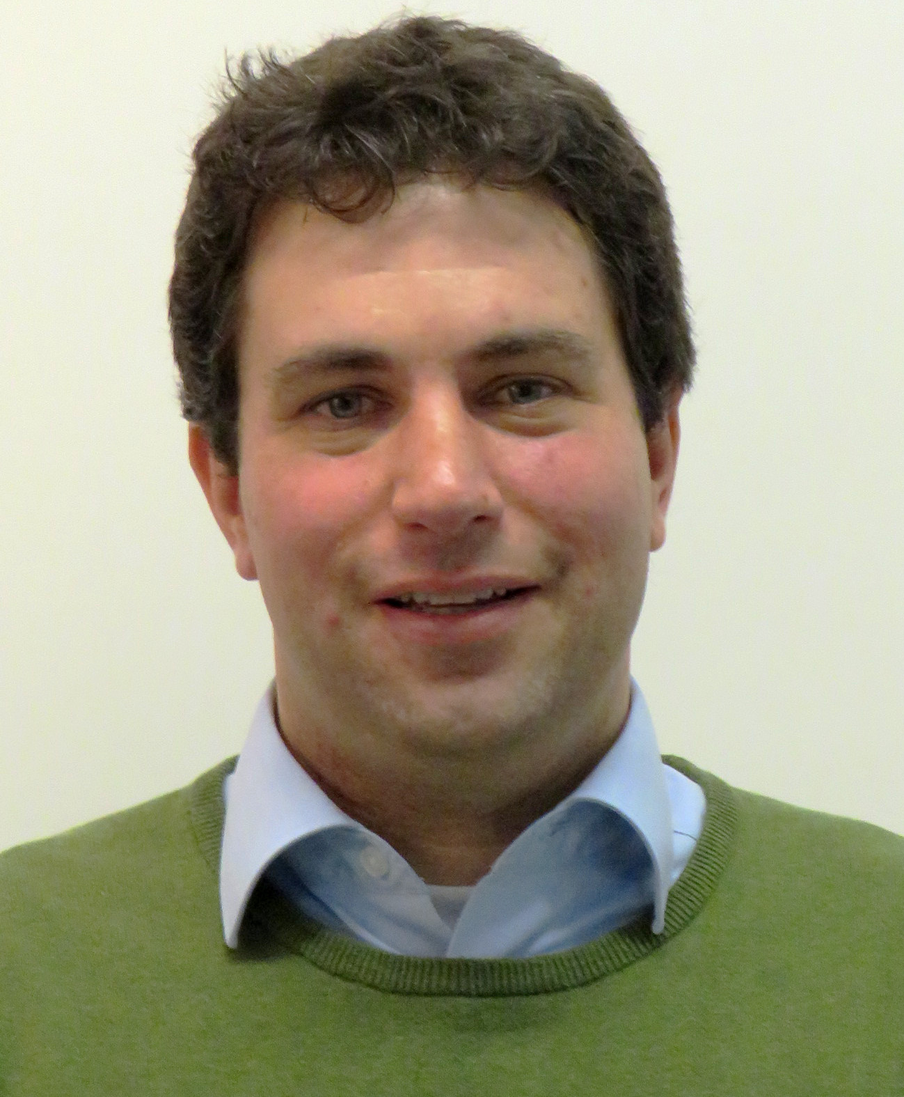
- MS4. Jakub Matusik
-
AGH University of Science and Technology, Krakow, Poland
-
Unraveling the structure-property relationship of adsorbents and photocatalysts derived from 2D layered materials of natural and synthetic origin -
Abstract: The materials which meet the requirements of several applications can be directly obtained from numerous geological deposits available worldwide. One example are clay minerals which are aluminum silicates having a 2D layered structure which is susceptible for chemical modifications. The lecture will provide selected examples of functional materials derived from clay minerals (mainly kaolin group minerals) as well as synthetic layered double hydroxides (LDH). In particular the presentation will focus on mesoporous nanotubular particles which can be derived from natural kaolinite mineral. The synthesis approaches for nanotubes formation and their properties will be discussed with emphasis on characterization with microscopic techniques. Finally the potential applications of kaolinite nanotubes will be presented with focus put on their loading with semiconductors (e.g. TiO2) for photodegradation of pollutants currently investigated by our research group.
-
Short bio: Prof. Jakub Matusik is currently employed at the AGH University of Science and Technology in Krakow (Poland). He obtained his PhD in 2010, habilitation in 2015 and graduated as full professor in 2021. By education, he is a geologist (clay mineralogist) working on the border of applied mineralogy and materials science. He has experience in designing, obtaining and testing nanomaterials based on various minerals. In particular he focuses on layered structures including natural clay minerals and synthetic layered double hydroxides as well as zeolites. Most of his research is devoted to adsorption phenomena and wastewater treatment. Other research areas include synthesis of clay-polymer nanocomposites and mineral-based (photo)catalysts. He leads a Mineral-based Architectures Group at AGH University: www.mba-group.agh.edu.pl
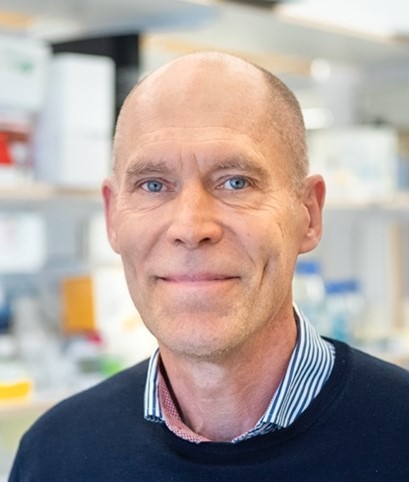
- LS4. Christer Betsholtz
-
Uppsala University & Karolinska Institute, Stockholm
-
New insights into the molecular and cellular composition of the brain barriers
-
Abstract: The brain is protected by both physical and biological barriers. The brain’s blood vessel walls are equipped with a blood-brain barrier, and the choroid plexus and the meninges are equipped with blood-cerebrospinal fluid barriers. To ensure that the brain is reached by wanted molecules, such as glucose, at the same time as it is protected by potentially neurotoxic substances circulating in the blood, the cells that form the barriers are equipped with efficient and specific molecular transporters. To learn more about
these barriers, we have analyzed the gene expression in the barriers’ cell types at the level of single cells using a technique referred to as single-cell RNA sequencing (scRNAseq). We identified distinguishing markers for individual barrier-forming cells types and subtypes, as well as other neighboring cell types, and used the markers to map the position of the cells in in vivo. I will present work in which we combined scRNAseq with immunofluorescence, in situ hybridization, transmission electron microscopy and immuno-EM to arrive at highly granular molecular anatomic maps of the brain barriers. This work revealed a surprising degree of cellular and molecular complexity and sophistication of the meninges, in particular the barrier located within the arachnoid mater.
-
Short bio: Betsholtz is currently a Professor at Uppsala University and Karolinska Institute. His research field of interest is vascular biology, in which he studies how cells communicate with each other and its surroundings when blood vessels form, and when they are affected by disease. He became Assistant Professor (1986) and Associate Professor (1990) at the Dept. of Pathology, Uppsala University. In 1994 he was appointed full Professor of Medical and Physiological Chemistry at Göteborg University, where he stayed until 2004, when he took a position as full Professor at the Dept. of Medical Biochemistry and Biophysics at Karolinska Institute, Stockholm. At 2013 he again took a new position, now as full Professor at Uppsala University. Between 2017 and 2020, he was Director of the Integrated Cardiometabolic Center at Karolinska Institutet. Today, he shares his time between Uppsala University and Karolinska Institute as Professor of Vascular Biology at both institutions. He has received various prestige awards such as the Louis-Jeantet Prize in Medicine, the Anders Jahre Prize in Medicine and the Fernström Nordic Prize. He is a member of the Nobel Assembly at Karolinska Institutet and member of the Swedish Royal Academy of Sciences. He received his Bachelor of Medicine degree from University of Uppsala in 1981, and his PhD degree in 1986. He was appointed Docent in 1987.
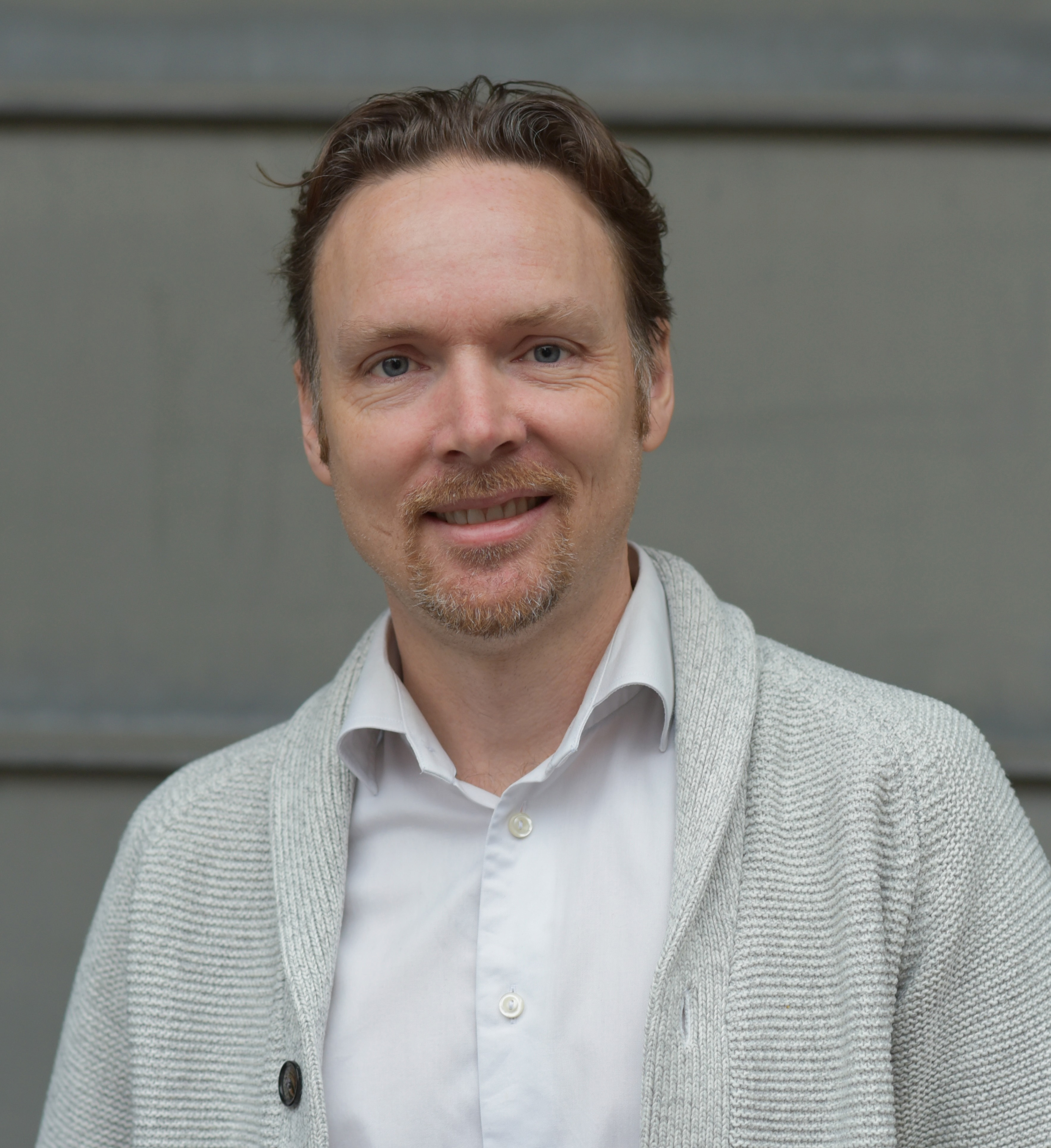
- MS5. Jani Kotakoski
-
University of Vienna, Austria
-
In and ex situ (S)TEM manipulation of 2D materials
-
Abstract: Structural engineering is the first step toward changing properties of materials, but has remained a challenge for 2D materials. The difficulties range from the preparation of clean and uniform samples to the sensitivity of these structures to the overwhelming task of sample-wide characterization of the subjected modifications at the atomic scale. I will demonstrate how these issues can be overcome using a near ultrahigh vacuum system comprised of an aberration-corrected scanning transmission electron microscope and setups for sample cleaning and manipulation, which are combined with automated atomic-resolution imaging of large sample areas and a convolutional neural network approach for image analysis. This combination of methods allows creating and fully characterizing atomically clean 2D materials, that can be further manipulated through introduction of defects and impurity atoms at the atomic scale.
-
Short bio: Jani Kotakoski has pioneered the research in atomic-scale structural manipulation of low-dimensional materials using electron and ion irradiation combining experimental materials physics, atomic-resolution transmission electron microscopy, and computational physics. He received his PhD in 2007 from University of Helsinki. After research periods at TU Darmstadt and University of Helsinki, he moved in 2011 to University of Vienna, where he is currently a full professor in condensed matter physics. His current research aims at creating atomically tailored structures embedded in solid state matrices to pave way for the second quantum revolution, and green energy production and storage.
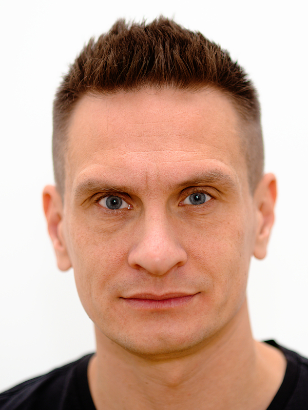
- LS5. Pontus Nordenfelt
-
Lund University, Sweden
-
Data-driven microscopy allows for automated context-specific acquisition of high-fidelity image data
-
Abstract: Light microscopy is a powerful single-cell technique that allows for quantitative spatial information at subcellular resolution. However, unlike flow cytometry and single-cell sequencing techniques, microscopy has issues achieving high-quality population-wide sample characterization while maintaining high resolution. I will present a general framework, data-driven microscopy (DDM), that uses real-time population-wide object characterization to enable data-driven high-fidelity imaging of relevant phenotypes based on the population context. DDM combines data-independent and data-dependent steps to synergistically enhance data acquired using different imaging modalities. We have developed plugins for improved high-content screening and live adaptive microscopy for cell migration and infection studies that capture events of interest, rare or common, with high precision and resolution.
-
Short bio: Pontus Nordenfelt is an Associate Professor and Senior Lecturer at Lund University. He is committed to a quantitative understanding of immunology, cell biology, and microbiology, with a primary interest in antibody function, infection processes,and cell migration.
He did his Ph.D. at Lund University and University of Toronto, focusing on molecular and cell biology of phagocytosis of bacteria. This was followed by a postdoc at Harvard Medical School, partly at MBL Woods Hole, with a focus on integrin biology and cell migration. He was also a fellow with the Image and Data analysis core at Harvard. After starting his own lab at Lund University, he has continued to develop microscopy and image analysis technologies and applied these to research into antibody function, infection, and cell migration. Technologies and molecules of interest include but are not limited to quantitative microscopy, live imaging, single-cell studies, antibodies, complement, and integrins.
- MS6. Demie Kepaptsoglou
-
University of York, UK
-
Advances in high energy and spatial resolution STEM EELS
-
Abstract: Engineering the structural or chemical architecture of functional materials at the nano or even atomic level enables emergent properties that rely on the interplay between fundamental properties of matter such as charge, spin and local atomic-scale chemistry. Thanks to advances in monochromators, state-of-the-art electron energy loss spectroscopy (EELS) in the scanning transmission electron microscope (STEM), offers nowadays the ability to map materials and atomic structures with an angstrom size electron beam and an energy resolution for EELS under 5meV. These capabilities have allowed to probe the spectroscopic signature of phonons down to the single atom level. Here, we present strategies for high spatial and energy resolution STEM-EELS experiments to interrogate proximity effects of heterostructure materials at the atomic scale and present opportunities for new experiments using monochromated electron probes.
-
Short bio: Demie Kepaptsoglou is a Staff Scientist of the SuperSTEM Laboratory in Daresbury UK and holds a joint Senior Lecturers position at the University of York. She received her PhD on Metallurgy and Materials Science from the National Technical University of Athens, Greece and subsequently worked as a Postdoctoral Associate at the university of Oslo in Norway, before joining SuperSTEM in 2011 and the University of York in 2017. Her work focuses on the implementation of analytical electron microscopy and spectroscopy in functional materials focusing on the effects of the presence of defects in their electronic structure and transport properties.
- LS6. Anders Tengholm
-
Uppsala University, Sweden
-
Live-cell imaging of sub-membrane signalling and secretion
-
Abstract: Critical to the understanding of cell function is availability of methods for recordings of intracellular regulators in single, living cells. Many key cellular events take place in the immediate vicinity of the plasma membrane. This presentation will highlight imaging approaches for the investigation of sub-membrane signalling of importance for the secretion of blood-glucose-regulating hormones insulin and glucagon from beta- and alpha-cells in the pancreatic islets. It will be shown how nutrients and neurohormonal stimuli induce dynamic changes of intracellular ATP, calcium ions and other second messengers and downstream effector proteins in the islet cells to shape the hormone release patterns.
-
Short bio: Anders Tengholm is a professor at the Department of Medical Cell Biology at Uppsala university. He completed his PhD at Uppsala university in year 2000. Following a postdoc period at Stanford university, he returned to Uppsala, where he conducts research aiming at understanding intracellular signalling processes that control secretion of blood-glucose-regulating hormones and the mechanisms underlying defective hormone release in diabetes. Tengholm utilizes various optical sensors and live-cell imaging approaches to monitor changes in second messenger concentrations, localization and activity of signalling proteins and exocytosis in single cells and in the intact pancreatic islet organ.
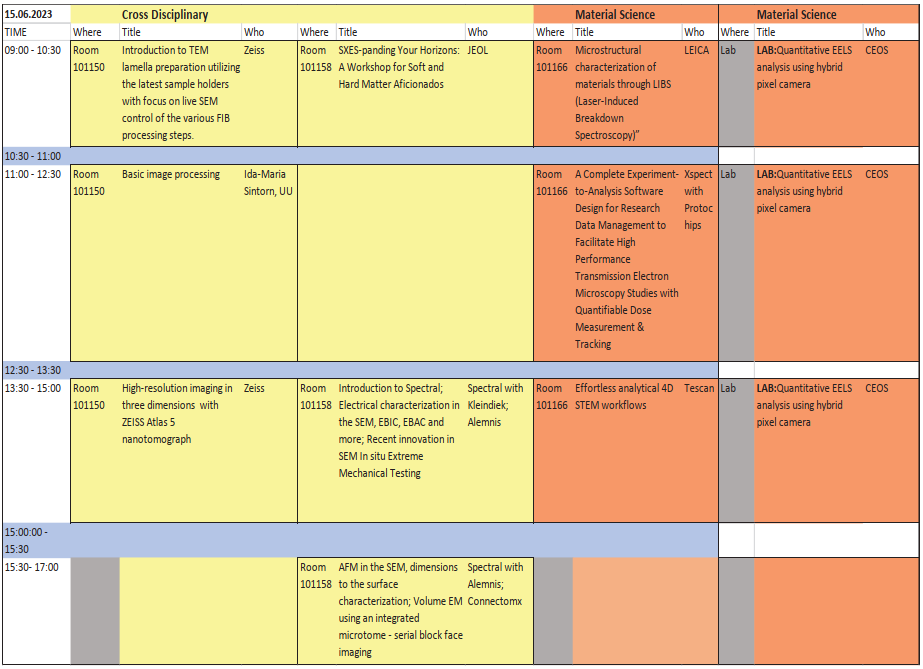
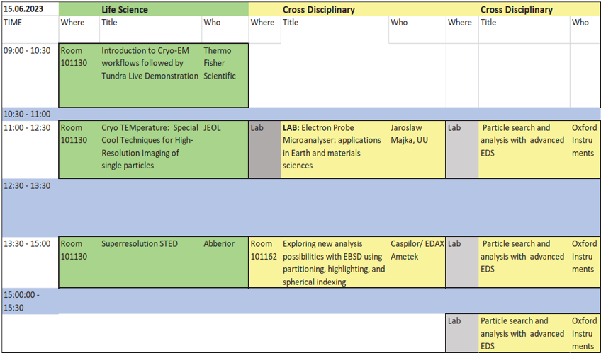
SCANDEM2023 offers a variety of workshops and labs from both academia and industry on Thursday June 15. Most workshops will be held in seminar rooms
in the same building where the conference takes place. For all laboratories, the meeting point is 10 minutes before the start of the laboratory at
Café Ångström. You will then be guided to the laboratory. The workshop program is shown below and is also found in the program booklet.
The SCANDEM2023 Conference welcomes companies to take the opportunity to represent their product portfolio in the SCANDEM 2023 exhibition area.
Meet your academic customers and present your products and solutions, raise awareness and interest in new developments in the fast-evolving field of microscopy.
Let us together find ways to improve current research and instrumentation.
An exhibitor invitation document can be downloaded
here (pdf)
House 10 at the Ångström laboratory
SCANDEM2023 will take place at House 10 at
the Ångström laboratory. This, the newest building at Uppsala University was inaugurated spring 2022. It hosts modern educational premises, learning environments and student spaces all designed to stimulate interdisciplinary approaches and new meetings within the University as well as cross-sector collaborations with industry and society. It is located approxaimtely 3km south of central Uppsala. The Ångström laboratory is easily reached by a short (~10min) busride from central Uppsala or by a beautiful 30 min stroll along the river and city garden.
Uppsala University
Uppsala University was founded in 1477 and is today a strong and broad research university. Here you will find 50,000 students and close to 5,000 researchers, spread over approximately 60 departments and similar units in three disciplinary domains: humanities and social sciences, medicine and pharmacy, and science and technology.
One of the University's famous alumni is Svante Pääbo, who was awarded the Nobel Prize in Physiology or Medicine 2022. Carl Linnaeus, Anders Celsius and Olof Rudbeck the Elder are a few examples of prominent scientists in
Uppsala University's history .
Uppsala
Uppsala is the fourth-largest city in Sweden, with close to 200 000 inhabitants. It is located 71 km (44 mi) north of the Swedish capital Stockholm. Since 1164, Uppsala has been the ecclesiastical centre of Sweden, being the seat of the Archbishop of the Church of Sweden. Uppsala is home to Scandinavia's largest cathedral - Uppsala Cathedral. The city hosts two universities: Uppsala university and the Swedish University of Agricultural Sciences.
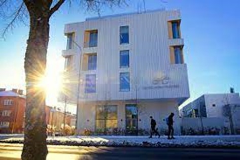
Hotel Von Kraemer,
https://hotelvonkraemer.se/
Conference rate per night with breakfast
Standard room: 1710 SEK/night
Check-in date: 11 Juni 2023
Check-out date: 15 Juni 2023
To book a room with the conference rate, please contact the hotel by email and reference the booking code:
SCANDEM
Phone: 018-495 99 00
Email:
info@hotelvonkraemer.se
The conference rate and rooms will be available until 11 Maj. Room reservation is made on a first come – first served basis.
Cancellation conditions: Double check with the hotel
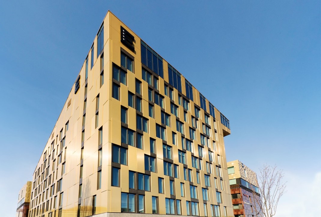
Elite Academia Uppsala,
https://elite.se/sv/hotell/uppsala/hotel-academia/
Conference rate per night in standard room with breakfast
sun-mon: 1450 SEK/night single room, 1550 SEK/night double room
Mon-wed: 1900 SEK/night single room, 2000 SEK/night double room
Check-in date: 11 Juni 2023
Check-out date: 15 Juni 2023
To book a room with the conference rate, please contact the hotel by email and reference the booking code:
SCANDEM
Phone: 018 780 99 00
Email:
info.academia@elite.se
The conference rate and rooms will be available until 11 May. Room reservation is made on a first come – first served basis.
Cancellation conditions: Double check with the hotel.
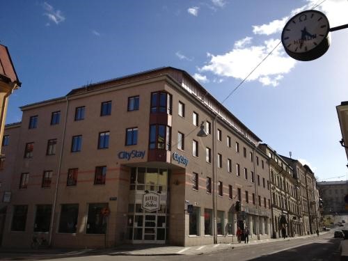
City Stay Uppsala,
http://citystayuppsala.se/
Conference rate per night with breakfast
Single economy room 1095 SEK/night
Single standard room 1295 SEK/night
Economy double room for singe use 1195 SEK/night
Standard double room for single use 1395 SEK /night
Check-in date: 11 Juni 2023
Check-out date: 15 Juni 2023
To book a room with the conference rate, please contact the hotel by email and reference the booking code:
SCANDEM23
Phone: 018 121 000
Email:
BOOKING@CITYSTAYUPPSALA.SE
Room reservation is made on a first come – first served basis.
Cancellation conditions: Double check with the hotel.