|
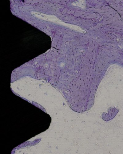
Until recently, implant integration was only evaluated from 2D microscopic
images. Reliable analysis of bone-implant integration requires 3D imaging as the whole sample should be included in the analysis.
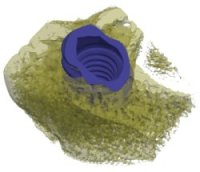
In this project we present two methods for visualization of SRμCT-scanned 3D volumes of screw-shaped bone implant samples: thread fly-through and 2D unfolding.
|
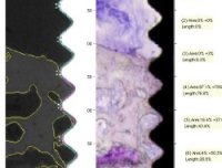
A necessary step for understanding the mechanisms of implant integration
is quantitative analysis of bone tissue around the implant. In this project, we present a segmentation method for the histological sections as well as SRμCT volumes and subsequent automatic quantitative analysis.
|
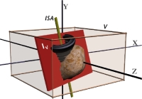
To enable a direct comparison between the two modalities
and to bypass the time consuming and difficult task of manual annotation
of the volumes, registration of these data types is desired.
In this project, we present two 2D-3D intermodal rigid-body image registration
methods for the mentioned purpose. To speed up the process, part of the computations
are done on the Graphic Processing Unit (GPU).
|


